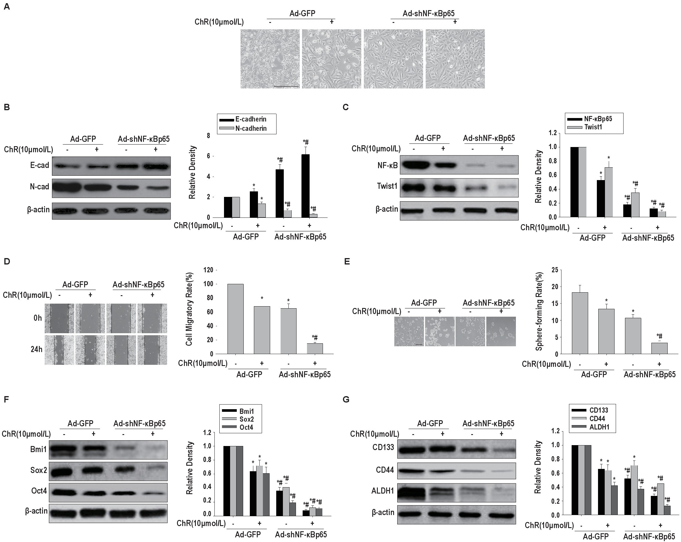
Fig. 3. Effects of NF-κBp65 knockdown on ChR associated inhibition of EMT and CSLC features in HeLa cells induced by TGF-β and TNF-α co-treatment. HeLa cells expressing shNF-κBp65 were co-administered TNF-α and TGF-β, with or without ChR(10.0μM). Then, cell morphology (A, scale bar, 50 μm), E-cadherin and N-cadherin protein levels (B), NF-κBp65 and Twist1 protein amounts (C), cell migration (D) and self-renewal (E, scale bar, 200 μm) abilities, and the protein expression levels of Bmi1, Sox2 and Oct4 (F), as well as CD133 and ALDH1 and CD44(G) were assessed. β-actin served as an internal reference for immunoblots. GFP, cells transduced with adenoviruses expressing GFP; shRNA, cells transduced with adenoviruses expressing shNF-κBp65. p<0.05 vs GFP;#p<0.05 vs GFP with ChR(10.0μM) treatment.